An international team of researchers at German Center for Neurodegenerative Diseases (DZNE) and Tokyo Institute of Technology (Tokyo Tech) have revealed in a collaborative study that neurons change on the molecular level when they are exposed to prolonged light. The researchers could identify that a feedback signalling mechanism is responsible for these changes. The innate neuronal property might be utilized to protect neurons from degeneration or cell death in the future.
Changes in the functional connections between neurons – 'synapses' – contribute to our ability to adapt to environmental changes. However until now, little was known about the signalling underlying such 'synaptic plasticity'. Now, investigations of fruit flies by researchers at DZNE, Tokyo Tech, the National Institute of Genetics in Japan, and the European Neuroscience Institute in Germany reveal details of the mechanisms behind synaptic plasticity.
Recent studies have suggested that changes in a region at the presynaptic membrane, described as the active zone, control synapse function. The research teams based in Germany and Japan exposed living fruit flies – the commonly studied Drosophila – to different light regimes and then compared the active zones in the photoreceptors.
T-shaped structures at the presynaptic membrane tether synaptic vesicles and control the release of neurotransmitters to the postsynaptic neuron. By tagging proteins that are crucial to these T-shaped structures the researchers revealed a drop in a subset of active zone proteins, while others remained unchanged. Further, they found that corresponding to the loss of structural proteins, the number of T-shaped structures was also reduced.
The researchers were also able to identify that a feedback mechanism was responsible for these changes and that it relied on the signalling protein Wnt. The results contribute to a better understanding of the molecular mechanisms underlying brain functions such as learning and memory. Future work may investigate how modifying the Wnt signal can be used to manipulate synaptic plasticity, with possible therapeutic applications for neurodegenerative or mental diseases.
"This synaptic change might reflect an innate neuronal property that leads to protection from excessive stimuli," explains Dr. Atsushi Sugie, the study's lead author. "By enhancing this property, we might be able to protect neurons from degeneration or cell death."
Background
Synapses
Synapses allow chemical signals to pass between neurons so that information can travel around the nervous system. Chemical signals are passed by the presynaptic release of neurotransmitters by synaptic vesicles. The neurotransmitters are received by postsynaptic receptors allowing the signal to pass from neuron to target neuron.
Repetitive or prolonged stimuli can lead to short- or long-term changes at synapses, affecting how they transmit signals and supporting plastic changes in the function of the nervous system, including learning and memory or the ability to adapt to environmental changes.
The role of proteins
Previous studies have identified that the probability of neurotransmitter release is strongly correlated with the levels of certain proteins at the active zone region of the pre-synaptic membrane. In addition, the localization of these proteins depends on neural activity. The present study reveals that the molecular composition of synapses can be modulated in living organisms exposed to sustained environmental stimuli.
Study details
Sugie and colleagues maintained Drosophila fruit flies that had just entered the adult stage of their life cycle in one of three light regimes: constant light; constant dark; and 12 hours light with 12 hours dark. The light used was mild to avoid retinal degeneration. Observations of fluorescently-tagged active zone protein Bruchpilot showed that while this protein was initially localised at discrete points representing active zones, prolonged exposure to light caused delocalization, which was reversed when the flies were returned to darkness.
A number of proteins have been associated with the structure of the active zone and the T-shaped structures that tether the synaptic vesicles. Although a subset of proteins was not affected, the researchers observed changes comparable to the loss of Bruchpilot in other proteins with similar functions, as well as re-organization of the microtubules.
According to the researchers the study suggests an accessible paradigm for assessing synaptic plasticity in neuronal synapses, as opposed to neuro-muscular synapses. They also emphasise how in their work they monitor synaptic plasticity with an optical microscope by tagging proteins with fluorescent molecules without the need for an electron microscope.
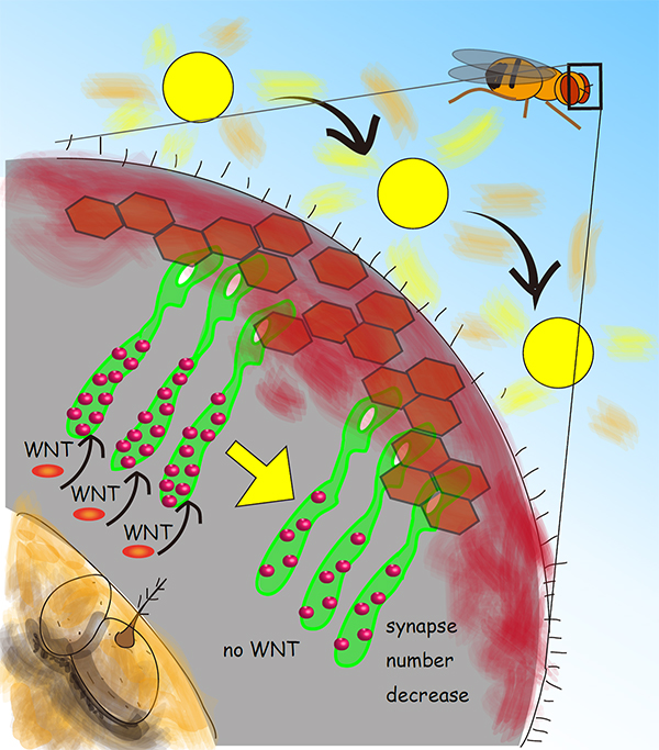
Fig. 1:
Schematic of the Drosophila visual system showing photoreceptor neurons (green) extending their axons to form synapses (red dots) with the brain neurons. When the fruit fly is exposed to natural light for 3 days the synapse number decreases. This suggests that the neuronal circuit plastically transforms by decreasing the number of efficient connections upon environmental changes, i.e. the light stimuli change. This research showed that the feedback signal mediated by the secreted protein WNT is required for the synapse change.
Source: Takashi Suzuki
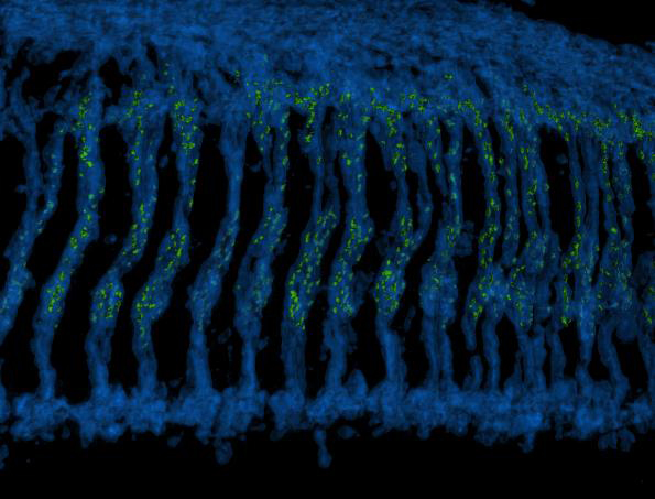
Fig. 2:
A 3D reconstructed image of the Drosophila optic ganglion where photoreceptor axons (blue) extend downwards to make connections with brain neurons. The synaptic connections formed by photoreceptors are highlighted with the active zone protein Bruchpilot and colored in green.
Source: Atsushi Sugie
Reference
Authors: |
Atsushi Sugie, Satoko Hakeda-Suzuki, Emiko Suzuki, Marion Silies, Mai Shimozono, Christoph Mohl, Takashi Suzuki* and Gaia Tavosanis*
*Co-corresponding authors |
Title of original paper: |
Molecular Remodelling of the Presynaptic Active Zone of Drosophila Photoreceptors via Activity-Dependent Feedback |
Journal, volume, pages and year: |
Neuron 86, 1-15, May 6, 2015 |
DOI: |
|