"Nanomachine Contrast Agent" using existing MRI detects minimal cancer tissue with a highly sensitive visualization
Tokyo Tech, the Innovation Center of Nanomedicine (iCONM), and the Japan Agency for Quantum and Radiological Science and Technology have developed a nanomachine contrast agent[1] which allows for a highly sensitive visualization of "hypoxic regions within the tumor"[2] using MRI. Such regions inside a tumor, even a microscopic environment, are malignant and resistant to treatment. The delivery of enough medicine to hypoxic regions inside a tumor is difficult and effects of radiotherapy are low. Since such regions show resistance to treatment, they are considered to be the regions which advance into more malignant forms of cancer and cause metastasis. The developed nanomachine contrast agent detects the microscopic environment of cancer tissue and has a previously unknown feature of amplifying MRI signal strength. The research team has shown that it makes possible tumor-specific imaging superior to existing MRI contrast agents. By using this nanomachine contrast agent, the team has also succeeded in using its high sensitivity to detect a small colorectal cancer only 1.5 mm in diameter which had metastasized to the liver. As described, the nanomachine contrast agent is minimally invasive compared to biopsy widely used in clinical settings. It is expected to be practically applied as "technology of pathological diagnosis by imaging" to many organs and tissues of the body. The nanomachine contrast agent can also be applied in treatment, for predicting the effects before treatment, or for a prompt effect evaluation afterward. In the future, it is expected to make possible reliable diagnoses that do not miss cancers and proactive treatment which is more reliable.
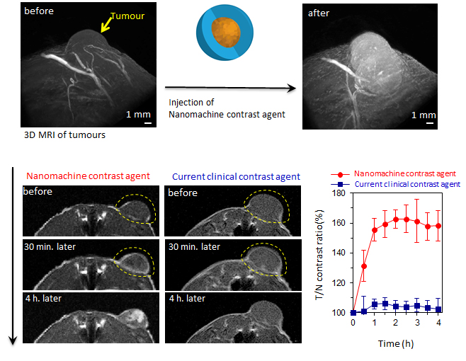
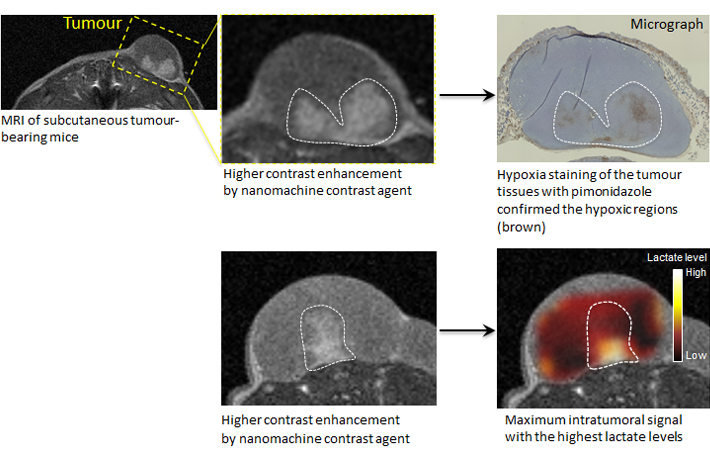
Figure 1. The nanomachine contrast agent makes possible the detection of cancer with a higher contrast than current contrast agents. Further, the regions of higher malignancy inside the tumor mass-produce more intense signals, providing additional information about the structure and the characteristics inside the tumor mass.
Figure 2. The nanomachine contrast agent may not only detect cancer with an MRI but also be effective in diagnosing its internal structure and malignancy. The nanomachine contrast agent produced stronger signals and made white the regions of low oxygen concentration and low pH levels thought to be especially malignant, even among cancer tissue. This effect was stronger in low-cost MRIs using lower magnetic fields. This means that MRI equipment existing at clinical sites can be used, and this new technology is expected to aid in diagnosing the degree of malignancy of cancers and their resistance to treatment.
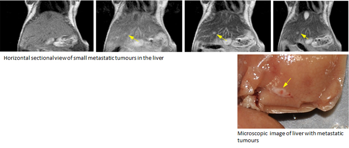
Figure 3. The nanomachine contrast agent produced a weaker signal in normal liver and a strong one in liver cancer, producing an extremely strong contrast. This enabled the detection of a small 1.5 mm colorectal cancer which had metastasized to the liver.
Reference
Authors: |
Peng Mi, Daisuke Kokuryo, Horacio Cabral, Hailiang Wu, Yasuko Terada, Tsuneo Saga, Ichio Aoki, Nobuhiro Nishiyama, Kazunori Kataoka |
Title of original paper: |
A pH-activatable nanoparticle with signal amplification capabilities for non-invasive imaging of tumour malignancy |
Journal: |
Nature Nanotechnology |
DOI : |
|
Explanations of Technical Terms
- [1]
- Nanomachine contrast agent
A nanomachine contrast agent of manganese contrast agent (which serves as an MRI contrast agent) confined within "calcium phosphate nanoparticles" which disintegrate in the low-pH environment of cancer tissue. This nanomachine contrast agent has an inner core containing a contrast agent surrounded by an outer shell of polymeric materials which are highly biocompatible. It is stable in the bloodstream environment (pH7.4) and releases manganese ions in response to the pH level inside a tumor (6.5-6.7). In addition, it has a property which amplifies the signal approximately seven times as the manganese ions released from the nanoparticle bind to proteins in cancer tissue and limit the movement of molecules. With these outcomes, it was shown that the nanomachine contrast agent has a characteristic of responding to slight changes in pH inside a tumor (6.5-6.7), causing the MRI signal to change.
- [2]
- Hypoxic regions within the tumor
Inside a malignant tumor arise regions of hypoxia, where not enough oxygen is supplied. Further, tumor blood vessels, which are built with a delay following growth of a tumor, are more fragile than those of normal tissue and are prone to repeated blockage or reflux. This is known to temporarily create a hypoxic environment, even in the vicinity of a tumor blood vessel. Cancer cells in such a hypoxic environment are reported to generally show resistance to anticancer drugs as well as radiotherapy. It is also said that cancer cells in hypoxic environments advance their migratory and motile abilities (their capacity for movement) to attempt to avoid poor environments, and this leads to the advancement of metastasis and the invasive capacity of the cancer.
- *
- This article has been updated to correct further information on June 16.