Researchers at Tokyo Institute of Technology (Tokyo Tech) have experimentally demonstrated a novel cancer diagnosis technique based on the scattering of circularly polarized light. Computational studies revealed that this technique can detect the progression of precancerous lesions and early cancer. This method can be implemented using an endoscope equipped with spin-LEDs—devices that emit circularly polarized light.
Most cancers of the digestive system emerge in the surface layer first and then progress into deeper layers. While surface layer carcinomas can be readily treated using an endoscope, carcinomas that have advanced onto deeper layers need surgical intervention to prevent them from metastasizing to lymph nodes or other organs. Thus, accurate measurements of the depth of cancer progression without damaging tissues are important to obtain useful information for making treatment-related decisions.
Current endoscopic diagnosis techniques like narrow-band imaging can only confirm the presence of cancer and distinguish between tumorous and non-tumorous tissue. There are very few direct measurement techniques that can provide a quantitative diagnosis of the depth and area of a carcinoma.
To tackle the abovementioned issue, a multinational research team led by Dr. Nozomi Nishizawa of Tokyo Tech recently conducted a study to demonstrate a novel cancer diagnosis technique using circularly polarized light. Their findings have been published in the Journal of Biophotonics , and a scientific illustration of the study was selected as an inside cover in the journal (Figure 1).
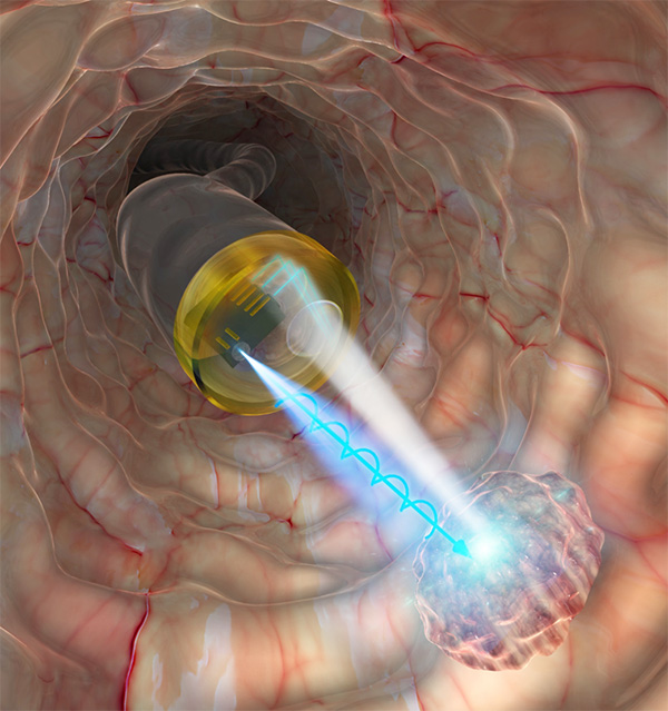
Figure 1. In vivo cancer diagnosis
Schematic illustration of the proposed technique using spin-LEDs on the tip of an endoscope. This image was selected as an inside cover of Journal of Biophotonics.
Their approach relies on how circularly polarized light interacts with healthy and unhealthy cells. "The depolarization of circularly polarized light scattered from biological tissues depends on structural changes in cell nuclei, which can provide valuable information for detecting cancer concealed in healthy tissues,"
explains Dr. Nishizawa. The team experimentally demonstrated this fact by shining near-infrared circularly polarized light on sliced tissue samples of murine liver containing metastatic lesions derived from intrasplenically injected human pancreatic cancer cells. They observed clear differences in the degree of circular polarization of the light scattered from the samples depending on the state of the biotissue, showing that cancer identification is possible with this technique (Figure 2).
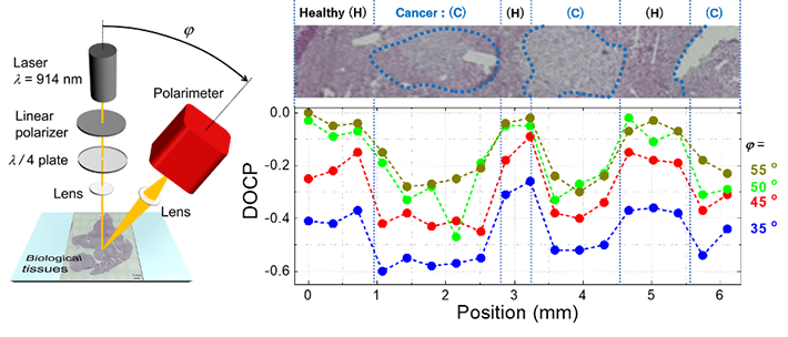
Figure 2. Experimental demonstration of the cancer diagnosis technique in biological tissue
- (left)Schematic illustration of the experimental setup. A laser beam with a wavelength of 914 nm is right-handed circularly polarized using optical filters and then focused on the sample. The scattered light was detected at an emission angle of φ.
- (right)Experimental results of line-scanning along the line which crosses the boundary between cancerous and healthy areas multiple times. The upper panel shows a microscopic view with cancerous parts surrounded by blue dotted lines. The lower graph shows the position dependence of the circular polarization of scattered light, which varied according to the state of the tissue.
Moreover, through computational studies with numerical simulations incorporating the scattering phenomena of circularly polarized light, the team also demonstrated that the depth profile of biotissues can be obtained by manipulating the detection angle. In short, the sampling depth in the target biotissue becomes deeper as the emission angle of the scattered light becomes close to perpendicular. Therefore, this dependence on the emission angle provides information on the depth profile of tissues or, in other words, the cancer’s progression toward the deeper layers.
However, one technical challenge had to be addressed to make this diagnosis method feasible: circularly polarized light cannot travel through optical fiber without losing its polarization. Therefore, the use of circularly polarized light in vivo requires a compact source of circularly polarized light. One promising candidate for such a source is spin-LEDs—devices developed by the researchers. In 2017, they succeeded in creating spin-LEDs capable of emitting almost pure circularly polarized light at room temperature. "By combining our novel technique based on circularly polarized light scattering and spin-LED devices, we will be able to determine the progression of precancerous lesions in vivo," remarks Dr. Nishizawa. To this end, in their latest study, the team designed the structure of an endoscope probe containing circularly-polarized LEDs, which can detect scattered light with various emission angles simultaneously (Figure 3).
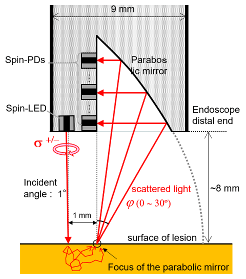
Figure 3. Cross-section of the designed endoscope probe for in vivo diagnosis of cancer progression
Figure 3. Cross-section of the designed endoscope probe for in vivo diagnosis of cancer progression
The probe consists of one spin-LED as an irradiation source, a parabolic mirror, and some spin-polarized photo-diodes (spin-PDs), which are detectors of circularly polarized light. Circularly polarized light is irradiated from the spin-LED onto the tissue. The scattered light beams are separated according to their emission angle by the parabolic mirror, and then the beams are separately detected by each spin-PD. Details are described in Nishizawa et al., JJAP 59, SEEG03 (2020).
The researchers are hopeful that the proposed technique will find application in the diagnosis of ulcerative colitis and alcoholic cirrhosis in future. Moreover, it could also be applied for the observation of engraftments in regenerative medicine and transplant surgery.
Reference
Authors : |
Nozomi Nishizawa1*, Bassam Al-Qadi2, Takahiro Kuchimaru3 |
Title of original paper : |
Angular optimization for cancer identification with circularly polarized light |
Journal : |
Journal of Biophotonics |
DOI : |
|
Affiliations : |
1Laboratory for Future Interdisciplinary Research and Technology, Tokyo Institute of Technology
2College of Engineering and Technology, Palestine Technical University-Kadoorie
3Center for Molecular Medicine, Jichi Medical University
|
* Corresponding author's email: nishizawa.n.ab@m.titech.ac.jp


