The ultimate goal of cancer diagnostics is to develop sensitive imaging techniques for reliable detection of tumor malignancy in the body. Scientists at Tokyo Institute of Technology have come close to achieving this goal by developing an injectable imaging probe that can specifically detect solid tumors based on the activity of hypoxia-inducible factor (HIF) regulated by the ubiquitin-proteasome system.
Tumor detection using targeted fluorescent imaging probes is a promising technology that takes advantage of specific molecular events occurring in cancer tissues. However, currently available probes that use this technology fail to maximize their specificity for tumors because of strong off-target signals, and thus, have limited ability to detect small tumors in a short timespan after systemic injection.
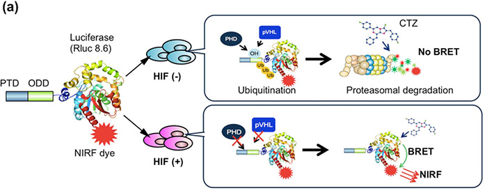
In order to overcome these problems, Dr. Kondoh and her colleagues at Tokyo Institute of Technology developed a novel, highly-specific, functional imaging probe that can detect hypoxic tumors with HIF activity, which is a hallmark of malignancy and poor prognosis. In normoxic cells, the transcription factor HIF-1α is degraded by the action of E3 ubiquitin ligase on the oxygen-dependent degradation domain (ODD) of HIF-1α; however HIF-1α is stabilized and therefore, accumulates in hypoxic cancer cells. Therefore, a protein containing the ODD would be intact in hypoxic tumors, but destroyed in normoxic tissues. The scientists exploited this molecular mechanism to construct a tumor-specific probe called POL-N (Protein transduction domain [PTD]-ODD-Luciferase-Near-infrared [NIR] dye) which contained: 1) the ODD core, 2) a PTD for intracellular delivery, 3) red-shifted luciferase, and 4) a NIR fluorescent dye with peak emission around 700 nm. This design enabled intramolecular bioluminescence resonance energy transfer (BRET) between luciferase and the NIR dye leading to generation of NIR light only in HIF-active hypoxic cells (Figure a). The normoxic cells did not generate BRET signals because POL-N was recognized through the ODD and degraded by the E3 ubiquitin ligase. Consequently, POL-N minimized the off-target signals and maximized tumor-to-normal tissue (T/N) ratio, while generating long-wave (700 nm) signals critical for imaging of deep tissues in living animals.
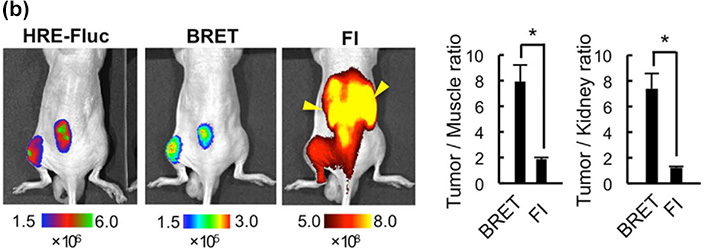
The scientists confirmed that POL-N-generated BRET signals correlated with hypoxia and intratumoral HIF activity, and successfully detected tumors in live mice, which was almost impossible using conventional fluorescence imaging (Figure b). Although the fluorescent dye detached from the degraded POL-N probe stayed for some time in normoxic cells, the intact POL-N probe rapidly disappeared from the circulation and excretory organs, including the kidney and liver, dramatically reducing off-target BRET signals because they are generated from only intact POL-N, and thus increasing tumor/muscle and tumor/kidney signal ratios (Figure c). Importantly, the injection of the POL-N probe enabled tracking of metastases in the liver and gastrointestinal tract, which are otherwise extremely difficult to detect by conventional fluorescence imaging because of strong off-target signals.
This research demonstrates that the technology combining BRET and ubiquitin proteasome system (UPS)-recognizable motifs is a breakthrough approach for fast and highly sensitive detection of tumors. Because the function of many important biological factors is controlled by the UPS, this study also offers a general strategy in the design of highly-specific injectable imaging probes for monitoring aberrant activity of the UPS-regulated factors both in cultured cells and whole organisms, thus opening new avenues in molecular imaging.


Figure. Injectable BRET imaging probe regulated by HIF-1α-specific UPS provided sensitive in vivo imaging of tumors. (a) A schematic diagram showing the behavior of the PTD-ODD-Luciferase-NIRF dye (POL-N) imaging probe in cells with (+) or without (-) HIF activity. CTZ, luciferase substrate coelenterazine. (b) Detection of subcutaneous tumors in mice using bioluminescence imaging with fire fly luciferase (HRE-Fluc), BRET probe imaging, and fluorescence imaging (FI) of the same mouse systemically injected with POL-N. Yellow arrowheads in FI indicate fluorescence signals from the kidneys. The image visualized using the BRET probe almost perfectly matches that of HIF-active tumor cells visualized using the HRE-Fluc reporter gene inserted into the tumor cell genome. (c) Tumor/muscle and tumor/kidney ratios of signals detected by BRET probe imaging and FI.
Reference
Authors: |
Takahiro Kuchimaru, Tomoya Suka, Keisuke Hirota, Tetsuya Kadonosono & Shinae Kizaka-Kondoh |
Title of original paper: |
A novel injectable BRET-based in vivo imaging probe for detecting the activity of hypoxia-inducible factor regulated by the ubiquitin-proteasome system |
Journal: |
Scientific Reports |
DOI : |
|
Affiliations : |
School of Life Science and Technology, Tokyo Institute of Technology |
- *
- This article has been updated to correct further information on October 25.
. Any information published on this site will be valid in relation to Science Tokyo.


