The protein collagen forms an integral part of our body's structures. However, there are gaps in our understanding of how the body produces collagen fibers. Looking to plug these gaps, Tokyo Tech researchers have developed a way to image the intracellular processing of a collagen precursor, type I procollagen, in real time! This could help us understand how defects in collagen formation could lead to diseases like liver fibrosis.
"Proteins are the building blocks of our body": You have probably heard this phrase before. Proteins are essential to the formation of our body’s structures, and their functioning.
Much like bricks and cement come together to form rooms, and then a whole building, multiple components come together to form the structure of our bodies. One of these components—a critical one, at that—is collagen. The fibers of this protein lay the foundation for our skin, muscles, bones, and any other organs in the body. It is on this foundation that our connective tissue is built!
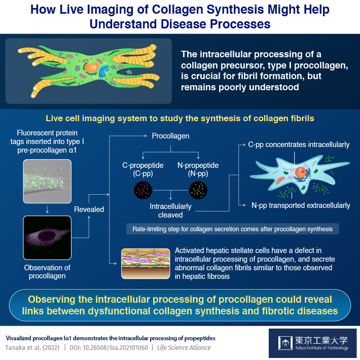
However, despite its importance in our body's structure, there were still some gaps in our understanding of how collagen fibers are synthesized by fibroblasts (which are cells specialized for this function). At this point, a group of researchers from Japan stepped in to take up the challenge. The team, led by Assistant Professor Dr. Toshiaki Tanaka of Tokyo Institute of Technology (Tokyo Tech), developed a live imaging system to observe how a specific type of procollagen—type I—is processed inside cells. Dr. Tanaka explains how they "Inserted fluorescent protein tags into type I pre-procollagen α1."
The tagging of procollagen (molecules that are formed inside fibroblasts and then processed into collagen) was previously discouraged, because addition of these tags compromised the delicate structure of collagen and resulted in abnormal collagen production. However, in their paper (which was published online on 18 February 2022, and then in Volume 5, Issue 5 of Life Science Alliance in May 2022), Dr. Tanaka's team mentions how they were able to identify regions where protein tags could be inserted without compromising collagen's structural stability. They then introduced this tagged procollagen into fibroblasts and studied them in real time, using confocal microscopy, immunostaining, and collagen assays.
What did they find? Dr. Tanaka tells us: "Fibroblasts are believed to secrete a collagen precursor—which consists of collagen, N-propeptide (or N-pp) and C-propeptide (or C-pp). Extracellularly, N-pp and C-pp are then cleaved by enzymes known as peptidases and go on to form collagen. But we found that these propeptides are cleaved inside the fibroblast." Live imaging studies showed that after being cleaved, N-pp was transported outside the cell, but C-pp accumulated around the nucleus of the cell, where it was further processed and degraded, before a small amount was secreted outside.
The intracellular processing, transportation, and secretion of collagen was hence identified as the slowest step in collagen synthesis. Understandably, the speed of this processing controls the speed of collagen synthesis. So, by monitoring this step with live imaging (as was done in this study), researchers can efficiently and quickly detect relative changes in collagen secretion!
In fact, the team went one step ahead and tried to detect these changes as well, specifically in liver fibrosis, a disease arising from the imbalanced secretion of collagen and certain other proteins by hepatic stellate cells (HSCs). This imbalance causes the formation of scar tissue in our liver, greatly compromising its normal functioning. The team used HSCs, which enable healing and injury response in our liver, for these experiments. Dr. Tanaka breaks it down for us, "In liver fibrosis, HSCs get activated and start secreting abnormally thick and long collagen fibrils. In our experiments, we observed that the production of such collagen fibers was linked to defective intracellular processing of procollagen."
In the future, Dr. Tanaka predicts the development of molecules and therapies to regulate collagen production. "These molecules could help treat diseases arising from an underlying disorder of collagen synthesis, like liver fibrosis."