Lipid membranes are more than just simple barriers separating cells and organelles from their surrounding environment. They also play key roles in several cellular functions, such as cell movement, material exchange, waste management, and sensing. In general, lipid membranes accomplish these feats with the help of proteins and other molecules, which are intricately integrated into the membrane structure, often modifying its fluidity or order. Accordingly, the study of lipid membrane order is an important subfield in cellular biology, not least because many diseases can cause or be caused by abnormalities in lipid membrane order.
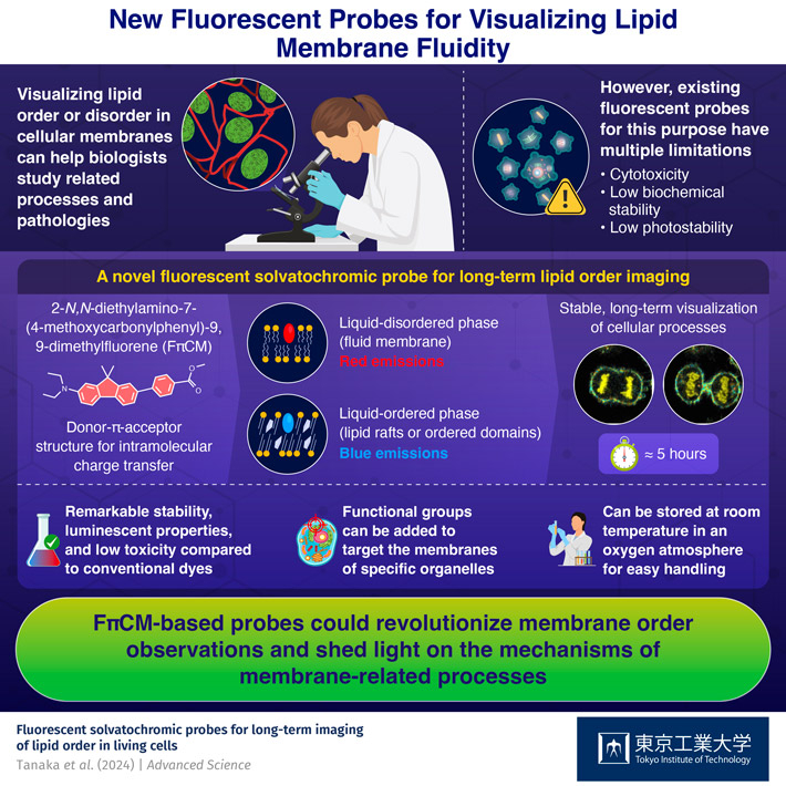
To visualize lipid membrane fluidity, scientists typically employ fluorescent substances known as solvatochromic probes or dyes. The term "solvatochromic" means that the light emitted by the molecule changes color depending on the polarity of the surrounding environment. Thus, when introduced into a lipid membrane, the color emitted by these dyes depends on lipid membrane order, which is closely related to polarity. However, conventional solvatochromic dyes face several challenges, including low stability, low fluorescent emissions, cell toxicity, and a reliance on ultraviolet light as an excitation source.
In a recent study published in Advanced Science (DOI: 10.1002/advs.202309721), a research team from Tokyo Institute of Technology and Kyushu University, Japan, sought to overcome all these hurdles. The research group, led by Associate Professor Gen-ichi Konishi of Tokyo Tech and Professor Junichi Ikenouchi of Kyushu University, developed a novel solvatochromic dye that could revolutionize real-time lipid order imaging.
To develop their new probe, the team first investigated and compared the photophysical properties of several different dyes. After some trial and error, they settled on a particular molecular design that met all their expectations. The final version of the probe, 2-N,N-diethylamino-7-(4-methoxycarbonylphenyl)-9,9-dimethylfluorene (FπCM), comprised a planar structure consisting of an electron donor part and an electron acceptor part joined together by a π-bridge. This configuration facilitated intramolecular charge transfers, which are essential to define the solvatochromic and fluorescent properties of the molecule.
The researchers evaluated the performance of the proposed dye through a comprehensive series of experiments. FπCM demonstrated exceptional fluorescent properties and remarkable chemical stability not only in solvents and artificial lipid membranes but also in physiological conditions in living cells. One of the most attractive aspects of the proposed dye was its long-term photostability, as Dr. Konishi remarks: "In our experiments, FπCM could persist for approximately five hours, whereas Prodan and Laurdan, two well-established solvatochromic dyes, would be completely quenched in approximately 30 minutes. The fact that we used relatively intense confocal laser light suggests that FπCM would also be resistant to the intense light coming from various devices."
Notably, the team could successfully observe lipid membrane fluidity during the entire process of cell division, implying that FπCM is non-toxic, unlike other state-of-the-art solvatochromic dyes. Moreover, the proposed probe can be readily modified to produce FπCM derivatives targeting specific lipid membranes, such as those found in cellular organelles like mitochondria and the endoplasmic reticulum.
"We believe that investigating the correlation between membrane protein activation in response to stimuli and spatiotemporal membrane fluidity transitions will shed light on the mechanisms underlying diverse membrane functions," concludes Dr. Konishi. "Since live imaging with FπCM and organelle-specific derivatives can easily be performed with conventional confocal microscopes, membrane order could become a standard, widely accessible information source for cell biologists."
With any luck, the exceptional properties of FπCM will aid biologists in unlocking the secrets behind the inner workings of cells.