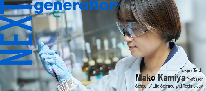
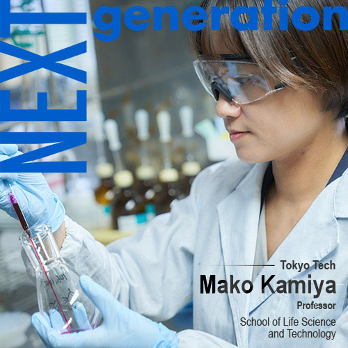
According to current estimates, one in two people develops cancer. One method of treating cancer is to surgically remove the cancerous tissue, but it is difficult to see cancer with the naked eye. To address this challenge, Professor Mako Kamiya of the Department of Life Science and Technology, School of Life Science and Technology, is working on a "fluorescent probe" that makes cancer glow. She is also a leading researcher in Raman probes, technology which is expected to dramatically increase the types of cancer cells that can be identified. Kamiya aims to help realize more precise cancer diagnoses and elucidate the mysteries of life processes and pathologies.
"Fluorescent probe" that makes cancer cells glow
- First of all, what kind of research do you do?
The three major standard treatment modalities for cancer are surgery, radiation therapy, and chemotherapy. Surgery is the removal of cancerous tissue. Surgery is believed to increase survival rates by removing as much of the residual cancerous lesion as possible. Therefore, not long ago, cancerous tissue and surrounding tissue were removed in large amounts. However, because of the adverse effects of removing normal tissue and the burden on the patient, methods to selectively remove cancerous tissue continue to be sought after.

However, it is not easy to remove only cancerous tissue. Although the location and size of the cancer is examined before surgery, during surgery it is difficult to detect with the naked eye where and how much cancer is present.
This led to the development of "intraoperative fluorescence imaging," the aim of which is to allow visualization of cancer cells during surgery by making them glow. In other words, a fluorescent substance is used to illuminate the site of a cancer or other lesion. This is expected to enable efficient surgical removal of cancerous tissue and eliminate omissions.
There is a global effort to develop "fluorescent probes," substances that make cancer cells glow. There are two types of fluorescent probes that make cancer glow: those that glow all the time, and those that glow only when they react with molecules characteristic of cancer cells. The latter is referred to as the "activatable type". We are focusing our research and development efforts on the activatable type because it has the advantage that even the smallest cancer lesions can be detected immediately.
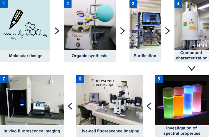
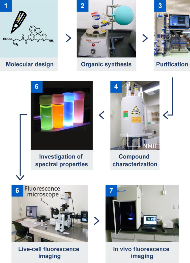
Research on probes at Kamiya Lab is conducted as follows. First, design the molecular structure of the probe according to an objective. Next, a compound is synthesized based on the design. Purify to extract only the desired compound from the synthesized product. In addition, nuclear magnetic resonance (NMR) and mass spectrometry (MS) are used for compound characterization. Then, evaluate whether it has the desired characteristics by spectral analysis. Finally, it is applied and administered to cancer cells or cancer model mice for imaging and confirmation. This cycle is repeated many times to obtain fluorescent probes with the desired characteristics.
- What are the characteristics of the fluorescent probes that you are developing?
We have focused on the enzymes of cancer cells and have been working on the research and development of fluorescent probes that glow only when they react with those enzymes. Before it reacts with a cancer enzyme, it does not glow even when exposed to light. But its special feature is that once it reacts with a cancer enzyme, it instantly glows brightly, allowing it to rapidly illuminate the cancer.
This project has been carried out in collaboration with Prof. Yasuteru Urano of the Graduate School of Pharmaceutical Sciences at the University of Tokyo and many lab members. When I first saw with the naked eye how the cancer in a mouse model of cancer glowed, I was convinced this would become a great innovation.
- What difficulties have you faced in researching fluorescent probes?
Since different types of cancer have different enzymes, it was difficult to find the optimal fluorescent probe. First, we increased the types of fluorescent probes and searched among these probes for one that glows from cancer. In doing so, we were assisted by clinicians from a variety of medical departments.
We were delighted when, as a result of our search, we found a promising fluorescent probe and were able to illuminate cancers in patient-derived specimens.
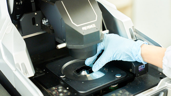
Fluorescence microscope installed in Kamiya Lab. Kamiya states that unlike conventional fluorescence microscopes, these microscopes are wonderful because they do not require a darkroom. When a fluorescent probe is set on a cell that has been made to allow cancer cells to glow, the cancer cell can be observed glowing.
Raman probe for simultaneous observation of multiple molecules in a cell
- What research are you focusing on now?
Since around 2018, in addition to fluorescent probes, we have begun development of an activatable Raman probe* that can be used in Raman imaging.
Raman imaging is an attempt to observe Raman signals originating from molecular vibrations using light. By detecting Raman scattering (one of the interactions between light and molecules) we can identify the type of molecule and analyze its molecular structure.
There are so many different types of molecules in the cell that interact with each other and function together. Therefore, a comprehensive understanding of life processes and etiologies requires simultaneous observation of more types of molecules and clarification of their interactions.
Fluorescence imaging has the problem of being limited to four or five types of molecules that can be detected simultaneously, while Raman imaging has the advantage of being able to detect more than 10 types of molecules simultaneously, surpassing fluorescence imaging.
- *
-
Raman probe: A generic term for chemical probes used in Raman imaging. By using Raman probes with different molecular vibration frequencies, multiple molecules can be detected simultaneously.
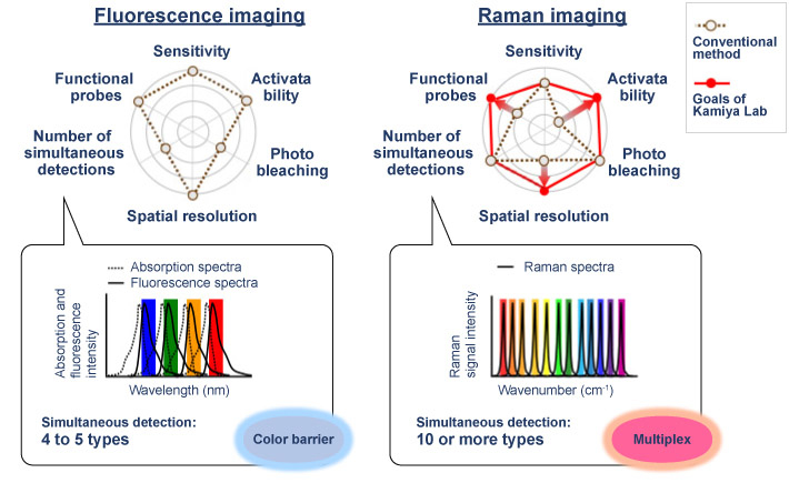
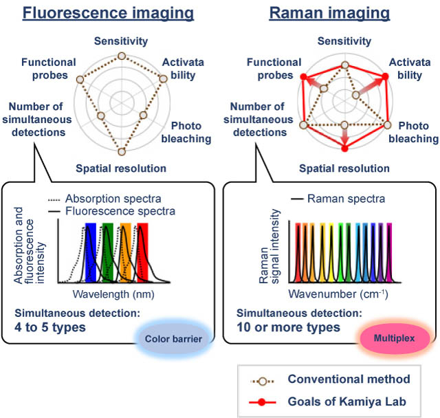
Compared to fluorescence imaging, Raman imaging has the advantage of being able to detect more types of molecules simultaneously.
- I see the importance of being able to see multiple types of molecules.
The type and combination of enzymes that each type of cancer has differ. Therefore, we believe that if we can observe more patterns of enzyme activity by using Raman probes that detect multiple enzyme activities, we will for example be able to make more precise diagnoses. It may also become possible to propose effective treatments according to the character of the cancer, such as a certain drug for a certain pattern of enzyme activity.
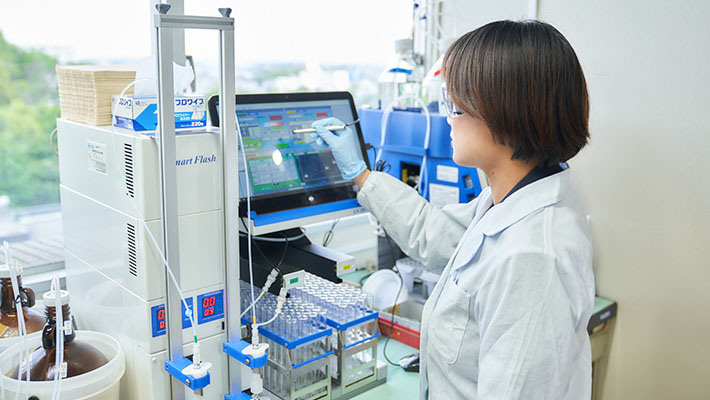
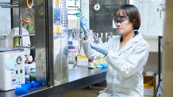
Kamiya purifying compounds that can be used as fluorescent probes or Raman probes
Successfully developed the world's first activatable Raman probe
- What challenges does Raman imaging present?
A major challenge for Raman imaging has been the limited repertoire of activatable probes that glow only when they react with specific enzymes and other molecules in the cell. Raman signals are derived from molecular vibrations; thus it was thought that it would be difficult to control the Raman signal intensity.
However, one thing led me to think that perhaps we could develop an activatable Raman probe that would turn on the Raman signal only when it reacted with the enzyme of a cancer cell, and so I decided to begin researching that.
Professor Yasuyuki Ozeki of the University of Tokyo's Research Center for Advanced Science and Technology heard my research presentation at a workshop I attended in the summer of 2018 and approached me. According to Prof. Ozeki, there is a paper that is currently being discussed in the field of Raman imaging, and the structure of the compound in the paper is very similar to the structure of the fluorescent probe I developed.
I began collaborating with Prof. Ozeki on the possibility of applying my fluorescent probe design to Raman imaging to create an activatable Raman probe.
Then, in 2020, we successfully developed the first activatable Raman probe. It was a challenge with no precedents, and it was a continuous process of trial and error, but thanks to the efforts of our lab’s Assistant Professor Hiroyoshi Fujioka, we were able to really move this project forward.
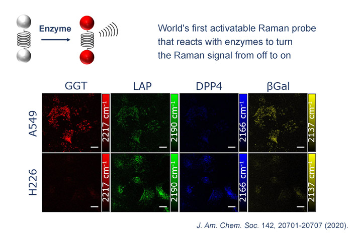
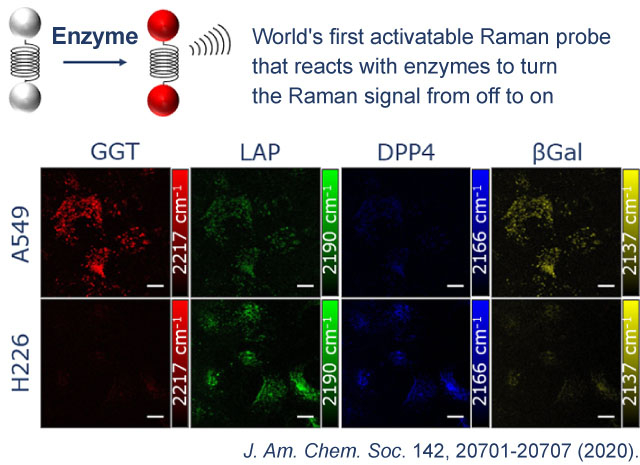
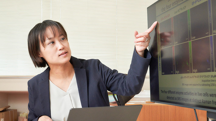
In 2020, in collaboration with Prof. Yasuyuki Ozeki of the University of Tokyo's Research Center for Advanced Science and Technology, Kamiya succeeded in developing an activatable Raman probe. Depicted here is enzyme activity in two lung cancers (A549 and H226) using four different Raman probes (GGT, LAP, DPP4, and βGal). Thus, hopefully having more types of enzymes and activity patterns that can be observed simultaneously will enable more precise cancer diagnosis.
- What factors made development of the probe possible?
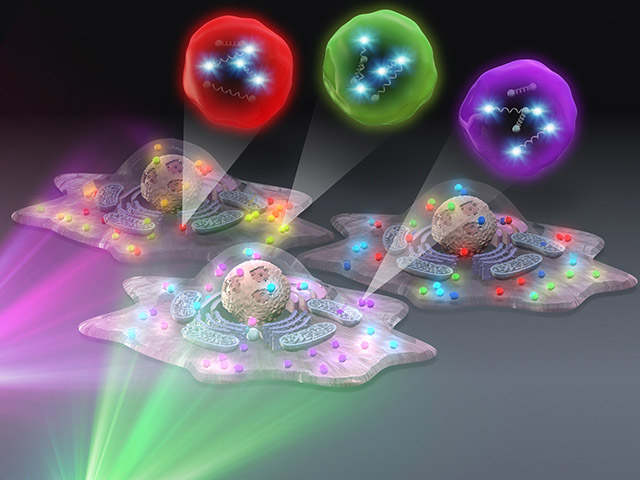
Concept image of Raman imaging, which glows in various colors in response to enzymes in cancer cells
I still think it is a matter of constantly being on the lookout to break new ground. Especially with regard to the activatable Raman probe, I feel that my efforts to collect information by actively participating in research groups and presenting my results have paid off.
- Please tell us about your future goals.
I would like to put the diagnostic and imaging methods using the newly developed fluorescent probes and Raman probes into practical use in clinical and research settings. I would be very happy if I could create something that biologists and physicians truly believe in, and if this could lead to the elucidation of the mysteries of life processes and etiologies.
Studying is the process of reading a textbook, and research is the process of creating a textbook.
- How did you become a probe researcher?

I enrolled at the University of Tokyo, and in my second year of undergraduate school, I had to choose a faculty. While visiting various faculties, I was attracted to and ended up choosing the Faculty of Pharmaceutical Sciences, which encompasses multiple fields such as biology, chemistry, and physics.
Furthermore, in my fourth year of undergraduate school, I was assigned to a laboratory. I chose Professor Tetsuo Nagano’s lab, working on fluorescent probes in the Laboratory of Chemistry and Biology, Graduate School of Pharmaceutical Sciences, The University of Tokyo. The reason I chose it was because when I went to visit the laboratory, I was impressed by the atmosphere and the fact that it is a domain that integrates biology and chemistry. In the laboratory, under the supervision of Prof. Nagano and Prof. Urano, and surrounded by great senior colleagues, junior colleagues, and friends, I had a wonderful research experience.
The fun of research on probes lies first and foremost in the thrill of creating entirely new compounds with your own hands. And if the synthesis is successful, one of the pleasures is to be able to observe its shiny glow. It was also a moving moment for me to see cancer cells that were invisible to the naked eye. I found myself turning into a researcher.
- What are your most important beliefs as a researcher?
Research can never be done alone. I always try to remember that I have many different people assisting me. I also want to create things that are not just self-satisfying, but that can actually be used in the real world by actively listening to the user's point of view and the needs of society. To this end, I value dialogue with those around me.
- In closing, do you have a message for students who want to become researchers?
I always tell students that studying is the process of reading a textbook, while research is the process of creating a textbook. Research is the process of revealing something previously unknown through experimentation and observation. Research is a continuous process of things going wrong and other difficulties. However, the moment you discover something new, or something that goes the way you want it to, is a very rewarding moment.
Nevertheless, it is hard to imagine what goes on in a research setting. Today, many universities, including Tokyo Tech, hold open campus events, so if there is research that interests you, I encourage you to take advantage of these opportunities to visit laboratories and talk directly with the researchers.
Mako Kamiya
Professor, Department of Life Science and Technology, School of Life Science and Technology
- 2022: Professor, Department of Life Science and Technology, School of Life Science and Technology, Tokyo Institute of Technology
- 2019: Associate Professor, Graduate School of Medicine, The University of Tokyo
- 2016: Lecturer, Graduate School of Medicine, The University of Tokyo
- 2014: Sakigake (PRESTO) researcher, Japan Science and Technology Agency (Concurrent post)
- 2010: Assistant Professor, Graduate School of Medicine, The University of Tokyo
- 2008: JSPS Research Fellowship for Young Scientists (SPD)
(Swiss Federal Institute of Technology Lausanne)
- 2008: Ph.D., Graduate School of Pharmaceutical Sciences, The University of Tokyo
- 2005: M.S., Graduate School of Pharmaceutical Sciences, The University of Tokyo
- 2003: B.S., Faculty of Pharmaceutical Sciences, The University of Tokyo

NEXT generation is a new series about emerging researchers, working at the forefront of science and technology as they envision the impact their discoveries will have on society's future.
The Special Topics component of the Tokyo Tech Website shines a spotlight on recent developments in research and education, achievements of its community members, and special events and news from the Institute.
Past features can be viewed in the Special Topics Gallery.